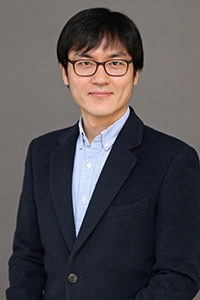
 Engineering Assistant Professor Jonghwan Lee is developing a new technique for the assessment of chemosensitivity in 3D culture, by maximizing the potential of a label-free 3D microscopy technology, called optical coherence tomography (OCT). He and co-investigators Diane Hoffman-Kim (associate professor of medical science and engineering), Jeffrey Morgan (professor of pathology and laboratory medicine and engineering), and Tao Liu (associate professor of biostatistics) are supported by a $1.8 million, five-year grant from the National Cancer Institute.
When selecting cancer therapy, physicians generally begin with first-line treatment options and monitor patient progress on a watch-and-wait basis, following a set of guidelines based on clinical trials from a large patient population. But this traditional method has been questioned on whether it provides individual patients with the optimal treatment.
To better find a matching treatment individually, a concept called precision cancer medicine, or personalized cancer medicine, has been studied. Among other approaches, functional precision medicine directly tests chemotherapy options on tumor cells biopsied from a patient to find the best matching treatment for the specific patient. This promising approach, however, has not been widely adopted by clinicians because the tumor microenvironment in a lab differed from the one within the patient’s body, leading to inconsistent drug responses between the sample and patient, and the quantity of biopsied cells was generally insufficient for a reliable number of options to be tested.
The first problem is being addressed by recent advances in three-dimensional (3D) cell culture techniques, which better mimic the body’s microenvironment in a lab. The second problem, the limited number of testable options, is mainly due to limitations in the current assay techniques that assess chemosensitivity in 3D culture. With most current assays, a sample can only be tested once, and multiple drugs with different mechanisms of action cannot be simultaneously tested by a single assay. Combined, these limitations exponentially reduce the number of testable options when involving multiple assessment time points to design a sequential therapy or when increasing the number of drugs to test a combination therapy.
“Because it is label-free, OCT was mainly used for investigating 3D morphology of objects in a specimen,” said Lee. “So I spent several years developing methods to extract more information from OCT images, not only by looking at 3D image patterns, but also by analyzing how the pixel values in an image vary with time when the sample is ‘alive’. I tested these new methods in 3D culture tissue, thanks to Jeff and Diane, and found they have a potential of assessing drug responses without tissue fixation, working as a kind of ‘live’ cell viability assay.”
Lee’s lab and the collaborators will use this new technique to image and analyze more than 100,000 spheroids for an unprecedentedly systemic investigation of the comprehensive range of OCT signal types.
“I believe the potential, when realized, will address the limitation of the current assay techniques and finally help to make the promising concept of functional precision cancer medicine available to a much larger population of cancer patients than currently possible,” he said.
This award is the fourth in six years for Lee from the National Institutes of Health, totaling about $7M.