Neuroscience Researchers Restore Leg Movement in Primates
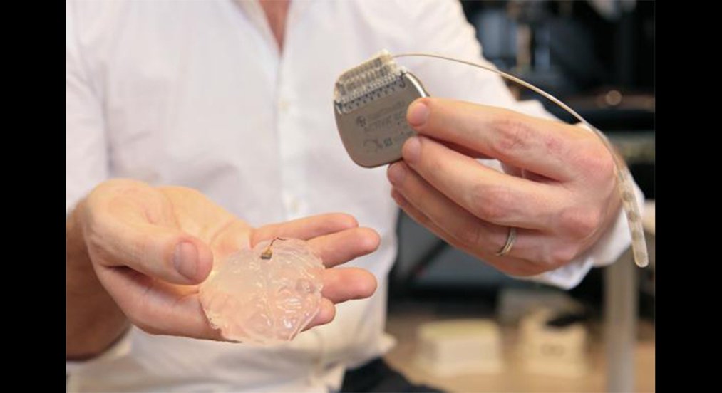
A Neural Bridge The brain-spine interface developed for this study uses a brain implant like this one to detect spiking activity in the brain's motor cortex. Seen here, a microelectrode array and a silicon model of a primate's brain, as well as a pulse generator used to stimulate electrodes implanted on the spinal cord. Credit: Alain Herzog/EPFL
The research is a step toward the development of a system that might help in rehabilitating people who have suffered spinal cord injuries.
An international team of scientists has used a wireless "brain-spinal interface" to bypass spinal cord injuries in a pair of rhesus macaques, restoring intentional walking movement to a temporarily paralyzed leg. The researchers, who describe their work in the journal Nature, say this is the first time a neural prosthetic has been used to restore walking movement directly to the legs of nonhuman primates.
The study was performed by scientists and neuroengineers in a collaboration led by Ecole Polytechnique Federale Lausanne (EPFL) in Switzerland, together with Brown University, Medtronic and Fraunhofer ICT-IMM in Germany. The work builds upon neural technologies developed at Brown and partner institutions, and was tested in collaboration with the University of Bordeaux, Motac Neuroscience and the Lausanne University Hospital.
"The system we have developed uses signals recorded from the motor cortex of the brain to trigger coordinated electrical stimulation of nerves in the spine that are responsible for locomotion," said David Borton, assistant professor of engineering at Brown and one of the study's co-lead authors. "With the system turned on, the animals in our study had nearly normal locomotion."
The work could help in developing a similar system designed for humans who have had spinal cord injuries.
"There is evidence to suggest that a brain-controlled spinal stimulation system may enhance rehabilitation after a spinal cord injury," Borton said. "This is a step toward further testing that possibility."
Grégoire Courtine, a professor at EPFL who led the collaboration, has started clinical trials in Switzerland to test the spine-part of the interface. He cautions: "There are many challenges ahead and it may take several years before all the components of this intervention can be tested in people."
Re-establishing communication
Walking is made possible by a complex interplay among neurons in the brain and spinal cord. Electrical signals originating in the brain's motor cortex travel down to the lumbar region in the lower spinal cord, where they activate motor neurons that coordinate the movement of muscles responsible for extending and flexing the leg.
Injury to the upper spine can cut off communication between the brain and lower spinal cord. Both the motor cortex and the spinal neurons may be fully functional, but they are unable to coordinate their activity. The goal of the study was to re-establish some of that communication.
The brain-spinal interface used a pill-sized electrode array implanted in the brain to record signals from the motor cortex. The sensor technology was developed in part for investigational use in humans by the BrainGate collaboration, a research team that includes Brown, Case Western Reserve University, Massachusetts General Hospital, the Providence VA Medical Center, and Stanford University. The technology is being used in ongoing pilot clinical trials, and was used previously in a study led by Brown neuroengineer Leigh Hochberg in which people with tetraplegia were able to operate a robotic arm simply by thinking about the movement of their own hand.David Borton (right) helped to develop the wireless neurosensor technology in the lab of Brown engineering professor Arto Nurmikko (left).: Mike Cohea /Brown University
A wireless neurosensor, developed in the neuroengineering lab of Brown professor Arto Nurmikko by a team that included Borton, sends the signals gathered by the brain chip wirelessly to a computer that decodes them and sends them wirelessly back to an electrical spinal stimulator implanted in the lumbar spine, below the area of injury. That electrical stimulation, delivered in patterns coordinated by the decoded brain, signals to the spinal nerves that control locomotion.
To calibrate the decoding of brain signals, the researchers implanted the brain sensor and wireless transmitter in healthy macaques. The signals relayed by the sensor could then be mapped onto the animals' leg movements. They showed that the decoder was able to accurately predict the brain states associated with extension and flexion of leg muscles.
The ability to transmit brain signals wirelessly was critical to this work, Borton said. Wired brain-sensing systems limit freedom of movement, which in turn limits the information researchers are able to gather about locomotion.
"Doing this wirelessly enables us to map the neural activity in normal contexts and during natural behavior," Borton said. "If we truly aim for neuroprosthetics that can someday be deployed to help human patients during activities of daily life, such untethered recording technologies will be critical."
The researchers combined their understanding of how brain signals influence locomotion with spinal maps, developed by Courtine's lab at EPFL, which identified the neural hotspots in the spine responsible for locomotor control. That enabled the team to identify the neural circuits that should be stimulated by the spinal implant.
With these pieces in place, the researchers then tested the entire system on two macaques with lesions that spanned half the spinal cord in their thoracic spine. Macaques with this type of injury generally regain functional control of the affected leg over a period of a month or so, the researchers said. The team tested their system in the weeks following the injury, when there was still no volitional control over the affected leg.
The study showed that with the system turned on, the animals began spontaneously moving their legs while walking on a treadmill. Kinematic comparisons with healthy controls showed that the lesioned macaques, with the aid of brain-controlled stimulation, were able to produce nearly normal locomotor patterns.
Limitations and future work
While demonstrating that the system works in a nonhuman primate is an important step, the researchers stressed that much more work must be done to begin testing the system in humans. They also pointed out several limitations in the study.
For instance, while the system used in this study successfully relayed signals from the brain to the spine, it lacks the ability to return sensory information to the brain. The team was also unable to test how much pressure the animals were able to apply to the affected leg. While it was clear that the limb was bearing some weight, it wasn't clear from this work how much.
"In a full translational study, we would want to do more quantification about how balanced the animal is during walking and measure the forces they're able to apply," Borton said.
Despite the limitations, the research sets the stage for future studies in primates and, at some point, potentially as a rehabilitation aid in humans.
"There's an adage in neuroscience that circuits that fire together wire together," Borton said. "The idea here is that by engaging the brain and the spinal cord together, we may be able to enhance the growth of circuits during rehabilitation. That's one of the major goals of this work and a goal of this field in general."
The research was funded by European Community's Seventh Framework Program (CP-IP 258654, NeuWALK), International Foundation for Research in Paraplegia Starting Grant from the European Research Council (ERC 261247, Walk Again), The Wyss Centre in Geneva Marie Curie Fellowship (331602, e-WALK), Marie Curie COFUND EPFL fellowships, Medtronic Morton Cure Paralysis Fund fellowship, NanoTera.ch Programme (SpineRepair), National Centre of Competence in Research in Robotics Sinergia program (CRSII3_160696), Sino-Swiss Science and Technology Cooperation (IZLCZ3_156331) and the Swiss National Science Foundation.
 David Borton (right) helped to develop the wireless neurosensor technology in the lab of Brown engineering professor Arto Nurmikko (left).: Mike Cohea /Brown University