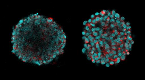
For Boutin's critieria, the ClearT2 method turned out to be clear winner. For her little balls of neural tissue – 100 millionths of a meter in diameter – ClearT2 allowed her to see fluorescing cells at all depths of focus and, importantly, it did not change the size of the tissue. Scale made her "neural spheres" substantially larger, while SeeDB made them smaller and didn't improve clarity as much.
Maintaining the tissue culture size is important because Boutin wants to know the physical dimensions of growth in the culture, such as the axons that one neuron might extend to another.
While ClearT2 worked within 1.5 hours, Scale and SeeDB took three days, Boutin said.
She ran several further tests with ClearT2, including with other types of healthy and cancerous neural cells, and ClearT2 continued to perform well, even allowing her to image extracellular matrix.
In the paper Boutin and Hoffman-Kim acknowledge that the results with different methods may vary with different samples, but they expect that for many "scaffold-free" engineered 3-D neural tissues – tissues that grow without added matrix supports – ClearT2 should work well.
Knowing that could clear the way for many research projects with 3-D tissues.
The National Science Foundation, The National Institutes of Health, and the Brown Institute for Brain Science supported the research. Imaging occurred in Brown's Leduc Bioimaging Facility. Boutin created her 3-D cultures with technology from Microtissues Inc., a company founded by Jeffrey Morgan, professor of medical science in the Department of Molecular Pharmacology, Physiology, and Biotechnology at Brown.
By David Orenstein